If you have ever had dental impressions taken the old-fashioned way, you probably remember it vividly. The cold, goopy material. The oversized tray pressed against the roof of your mouth. That moment where you wondered whether you might gag or whether the material would ever actually set. I took thousands of those impressions early in my career, and I can tell you honestly that I do not miss them one bit. Neither do my patients. Today, digital scanning has transformed this part of orthodontic treatment into something fast, comfortable, and remarkably precise.
What a Digital Scan Actually Involves
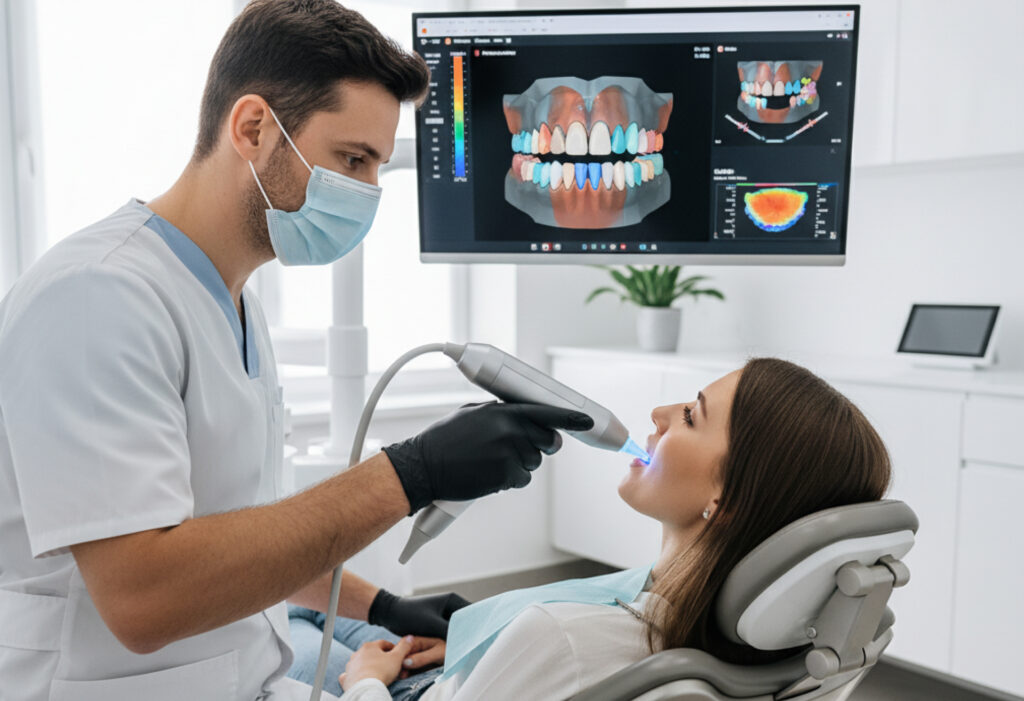
A digital scan for braces is a three-dimensional capture of your teeth, gums, and bite using a small handheld wand. The device uses optical technology, either laser or structured light, to record thousands of images per second as it passes over your dental surfaces. Software then stitches these images together into a highly detailed 3D model of your mouth. The entire process usually takes between two and five minutes, depending on how much of your mouth needs to be captured.
From the patient's perspective, it feels like someone is slowly moving a camera around the inside of your mouth. There is no material to bite into, no holding still while something hardens, and no unpleasant taste. You can breathe normally, swallow whenever you need to, and even pause if you feel uncomfortable. I have scanned patients as young as six and as old as eighty-two, and the universal feedback is that it is easy and painless.
How Digital Scans Compare to Traditional Molds
Are digital impressions better than traditional molds? In almost every measurable way, yes. Traditional impressions require a physical material, usually alginate or polyvinyl siloxane, to be loaded into a tray and pressed against the teeth until it sets. The material must be mixed to the right consistency, the tray must be positioned perfectly, and the patient must remain still for 60 to 90 seconds without gagging or moving their tongue. If anything goes wrong, you start over.
Digital scans eliminate all of these variables. There is no mixing, no trays, no setting time, and no risk of a bubble or tear ruining the impression. If the scanner misses a spot, the clinician simply goes back and rescans that area. The result is a model that is accurate to within 20 microns, which is far more precise than even the best physical impression.
I remember a teenage patient a few years back who had such a strong gag reflex that taking upper impressions was nearly impossible. We tried smaller trays, numbing spray, breathing techniques, everything. It took three attempts and she was in tears by the end. When we switched to digital scanning, the same area was captured in under two minutes with no discomfort at all. That experience alone convinced me that digital was the future.
The Technology Behind the Wand
Modern intraoral scanners use different technologies depending on the manufacturer, but they all accomplish the same goal. Some use confocal laser imaging, projecting a laser onto the tooth surface and measuring how the light returns to calculate depth and contour. Others use structured light, projecting a pattern of light onto the teeth and analyzing how that pattern distorts across different surfaces. Both approaches produce remarkably accurate results.
The software that accompanies these scanners is equally impressive. As the scan progresses, you can watch a full-color 3D model of your teeth build in real time on a screen beside the chair. Patients love seeing this because it gives them an immediate visual of their dental anatomy, often for the first time. I use these models to show patients exactly where their crowding is, how their bite fits together, and what we aim to achieve with treatment.
What Digital Scans Are Used For
In orthodontics, digital scans serve multiple purposes. They replace impressions for creating custom aligners, retainers, and certain types of braces. They allow us to digitally plan tooth movements and simulate end-of-treatment results before we ever begin. They provide a baseline record that we can compare against throughout treatment to monitor progress.
For patients receiving clear aligner therapy, the digital scan is the starting point for the entire treatment plan. The 3D model gets uploaded to specialized software where I can map out each stage of tooth movement. The patient can even see a simulation showing how their teeth will look at the end of treatment, all generated from that single scan.
Digital scans are also invaluable for creating retainers. When treatment ends, we scan the final result and have a retainer fabricated from that precise digital model. If a retainer ever breaks or gets lost years later, we can produce an exact replacement from the stored file without the patient needing to come in for a new impression.
Accuracy and Reliability
One concern patients sometimes raise is whether a digital scan is as reliable as the old-fashioned method. The research on this is clear: digital impressions are equal to or more accurate than traditional impressions in virtually all clinical situations. Multiple peer-reviewed studies have shown that the fit of appliances made from digital scans is excellent, often superior to those made from physical molds.
Part of the reason is that physical impressions introduce multiple opportunities for error. The impression material can warp, tear, or pull away from the tooth surface. It can be distorted during removal from the mouth. Stone models poured from impressions can chip or degrade over time. Digital scans bypass all of these failure points because the data exists as a file that never changes or degrades.
Patient Comfort and Accessibility
Beyond accuracy, the comfort factor is significant. Patients with strong gag reflexes, those with anxiety about dental procedures, and young children all benefit enormously from digital scanning. I have also found it helpful for patients with limited jaw opening, since the scanner wand is much smaller than a loaded impression tray.
The speed of digital scanning also means less time in the chair. A full-arch impression with traditional materials takes about four to five minutes of actual set time, plus preparation and cleanup. A digital scan of both arches and the bite typically takes three to four minutes total. For busy patients, and especially for fidgety kids, that time savings matters.
The Environmental and Practical Benefits
There is another advantage that rarely gets discussed: sustainability. Traditional impressions generate waste. The alginate or PVS material gets thrown away. The trays may be disposable. The stone models take up physical storage space and eventually end up discarded. Digital scans produce none of this waste. The files are stored on secure servers indefinitely, accessible whenever needed, without occupying a single inch of shelf space.
For orthodontic practices, this also means no more boxes of dusty plaster models in storage rooms. I used to have an entire closet dedicated to patient models. Now all of that information lives on a hard drive smaller than a paperback book.
Looking Ahead
Digital scanning technology continues to improve each year. Scanners are getting smaller, faster, and more affordable. The software is becoming smarter, with artificial intelligence helping to identify dental anatomy and flag potential issues. Some systems can now detect early signs of decay or gum recession during a routine orthodontic scan.
If you are about to start orthodontic treatment and you are dreading the idea of goopy impressions, ask your local orthodontist whether they use digital scanning. Most modern practices have adopted this technology, and those that have will tell you the same thing I tell my patients: the days of messy molds are behind us, and we are all better off for it.
Comments